 Wow! eBook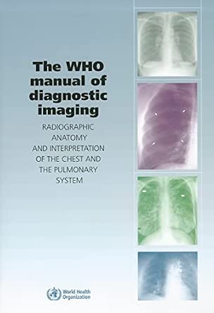
The WHO Manual of Diagnostic Imaging: Radiographic Anatomy and Interpretation of the Chest
Author(s): S.M. Ellis (Author), Christopher Flower (Author), Harald Ostensen (Editor), Holger Pettersson (Editor)
- Publisher: World Health Organization
- Publication Date: 3 Aug. 2006
- Edition: Illustrated
- Language: English
- Print length: 152 pages
- ISBN-10: 9241546778
- ISBN-13: 9789241546775
Book Description
The text aims to provide an aid to the interpretation of the chest radiograph (CXR). It is not a comprehensive account of all possible chest diseases but a descriptive text to help identify the way in which chest pathology is manifest and diagnosed on CXR. The initial chapters deal with interpretive skills and pattern recognition and the later chapters demonstrate specific pathologies.
Backed by high-quality reproduction of radiographs, this manual will prove essential reading to general practitioners, medical specialists, radiographers, and radiologists in any medical settings, although focusing specifically on needs in small and mid-size hospitals.


